
News|Articles|October 22, 2025
Fujifilm launches advanced endoscopy platform with new imaging capabilities
Author(s)Todd Shryock
Fact checked by: Chris Mazzolini
Listen
0:00 / 0:00
Key Takeaways
- The ELUXEO 8000 System enhances visualization and precision in gastrointestinal care with a 4K processor and advanced imaging modes, including Amber-Red Color Imaging.
- Compatible with existing Fujifilm endoscopes, the system supports new 800-series endoscopes with intelligent CMOS sensors for advanced diagnostic and therapeutic applications.
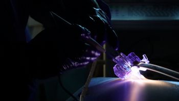
Fujifilm unveils the ELUXEO 8000 Endoscopic Imaging System, enhancing visualization and precision in gastrointestinal procedures with advanced imaging technology.
Advertisement
The ELUXEO 8000 System combines a 4K processor and light source in a single unit and features triple noise reduction, extended dynamic range, and new imaging modes intended to improve detection of abnormalities. Building on the company’s multi-light LED technology, the system introduces Amber-Red Color Imaging, designed to improve visualization of blood vessels and bleeding sources during complex procedures. It complements Fujifilm’s existing Linked Color Imaging and Blue Light Imaging modes, which support enhanced polyp detection and characterization.
“We are steadfast in our commitment to advance the field of endoscopy with innovations that empower GI physicians to safely and efficiently accomplish even the most challenging procedures with the best possible outcomes,” said Tai Fujita, vice president of the Endoscopy Division at FUJIFILM Healthcare Americas. “The ELUXEO 8000 System is our latest testament in how Fujifilm is taking endoscopic imaging to new heights.”
The platform is compatible with Fujifilm’s 500-, 600-, 700- and 800-series endoscopes, enabling facilities to upgrade imaging capabilities without replacing existing scopes. It also supports a new line of seven FDA-cleared 800-series endoscopes equipped with intelligent CMOS sensors for advanced diagnostic and therapeutic applications, including full access to the small intestine.
According to Hiroyuki Aihara, M.D., Ph.D., director of endoscopic tissue resection at Brigham and Women’s Hospital, “By emphasizing color contrast and structural detail, this new observation mode improves visual recognition of tissue layers and blood vessels when working in the submucosal space. Visualization of these structures is critical for safe and successful completion of ESD and other third space procedures.”
Recent advances in endoscopic imaging and therapeutic technology
The field of
Another major focus is the development of enhanced imaging modes that use specific wavelengths of light to detect vascular and structural changes below the surface of the mucosa. These digital and optical chromoendoscopy techniques are being increasingly used during procedures such as polyp detection, tissue resection, and third-space endoscopy. The use of targeted light modes improves visibility of blood flow and tissue layers, allowing clinicians to distinguish between benign and potentially malignant lesions more effectively.
Therapeutically,
Artificial intelligence is also emerging as a transformative tool in the endoscopy suite. AI-assisted detection algorithms can flag suspicious lesions in real time, reducing variability between operators and improving adenoma detection rates. Additionally, workflow automation and smart processors are helping clinicians streamline procedure protocols and documentation. As these technologies mature, endoscopy is transitioning from purely diagnostic to a fully integrated diagnostic-therapeutic discipline, supporting earlier intervention, reducing hospital stays, and enhancing long-term patient outcomes.
Advertisement
Related Content
Advertisement


Advertisement
Advertisement
Trending on Medical Economics
1
Medicare is pushing life-saving drugs out of physician offices — and older patients will suffer
2
The four forces reshaping physician pay, with Tynan Kugler of PYA
3
Medicare covers weight-loss drugs; one year of the One Big Beautiful Bill Act; American medicine at 250 — Morning Medical Update Weekly Recap
4
Your practice has more connected devices than ever. That's a cybersecurity problem
5